Gallery of Structures Solved in the CingolaniLab

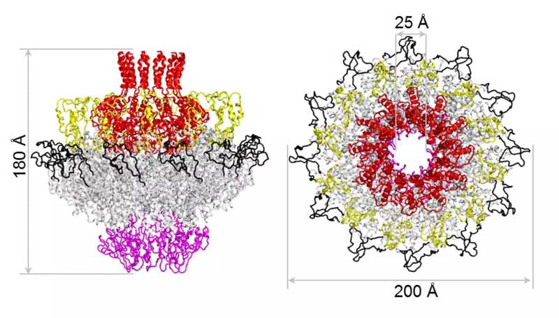
High-resolution cryo-EM analysis of the therapeutic Pseudomonas phage Pa223
PDB entries (Cryo-EM): 9NY2, 9NWI, 9NXK, 9NWM, 9NXO, 9NY6, and 9NXP
Maps: EMD-49916, EMD-49926, EMD-49882, EMD-49902, EMD-49887, EMD-49909, EMD-49925, and EMD-49910
Hou et al., J Mol Biol., 2025

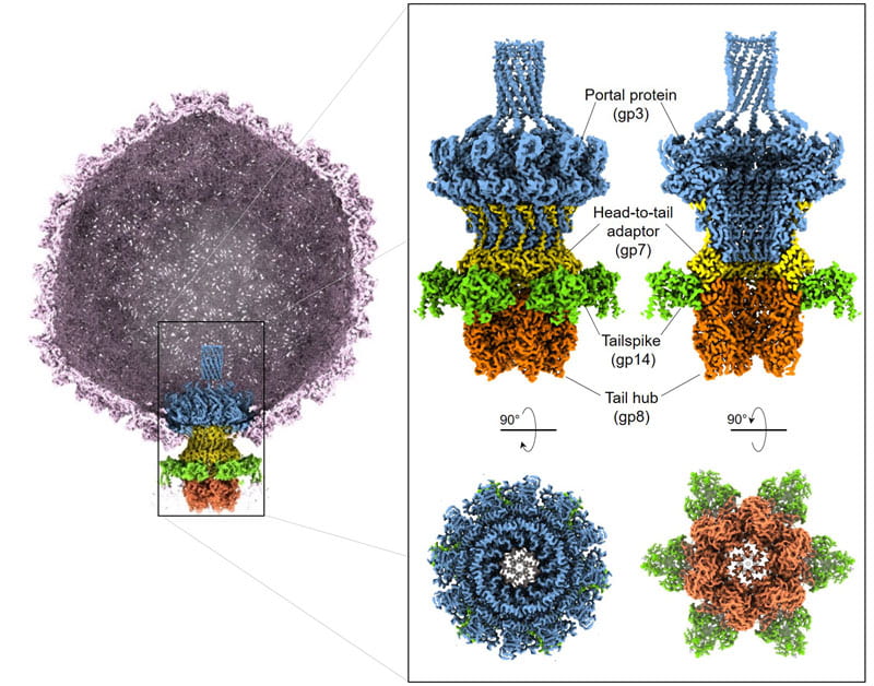
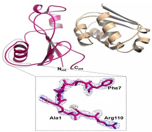
Structural atlas of the therapeutic Pseudomonas-phage Pa193
PDB entries (Cryo-EM): 9B40, 9B41, 9B42 and 9B45
Maps: EMD-44163, EMD-44164, EMD-44166, and EMD-44168
Iglesias et al., Commun Biol., 2024

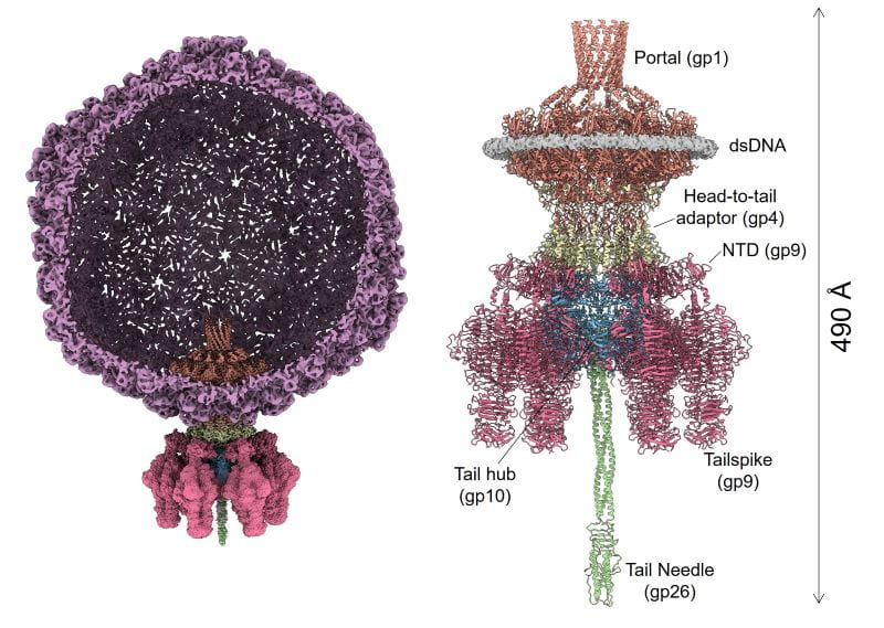
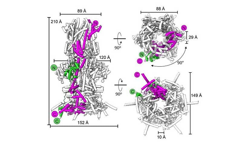
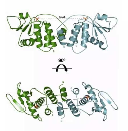
Architecture of Pseudomonas-phage DEV
PDB entries (Cryo-EM): 9BGN, 9BGM, 9BGO, 9COD and 8VXQ
Maps: EMD-44518, EMD-44517, EMD-44519, EMD-45776 and EMD-43629
Lokareddy et al., Nat Commun., 2024
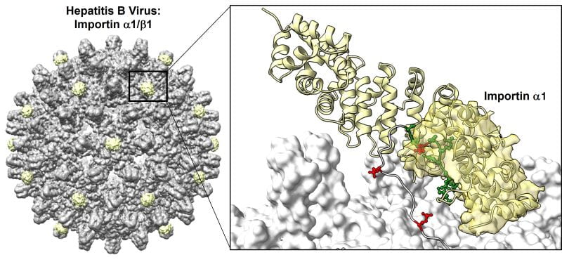
Hepatitis B virus (HBV) nucleocapsid bound to Importins
PDB entries (Cryo-EM): 8G5V, 8G8Y, 8G6V, and 8GCN
Maps: EMD-29756, EMD-29858, and EMD-29785
Yang et al., Science Adv., 2024
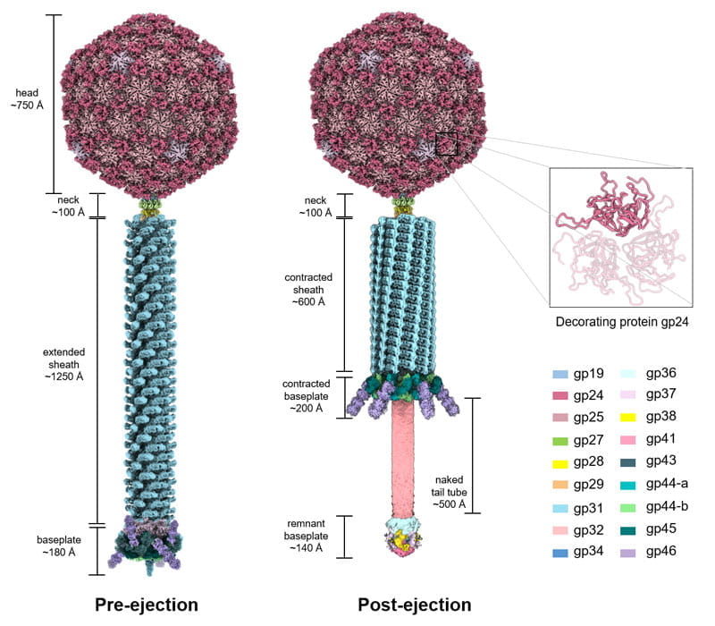
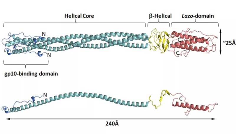
Salmonella-phage P22 mature virion
PDB entries (Cryo-EM): 8TVU, 8TVR, 8U10, 8U11, and 8U1O,
Maps: EMD-41651, EMD-41649, EMD-41791, EMD-41792, and EMD-41819
Iglesias et al., J. Mol Biol., 2023
Pseudomonas-phage E217
PDB entries (Cryo-EM): 8FRS, 8FVH, 8FUV, 8FVG, and 8EON,
Maps: EMD-29406, EMD-29487, EMD-29481, EMD-29486, and EMD-28405
Li et al., Nature Commun., 2023
Shigella-virus Sf6
PDB entries (Cryo-EM): 7SFS, 7SG7, 7SPU, 7SP4, and 7UKJ
Maps: EMD-25101, EMD-25106, EMD-23372, EMD-25365, and EMD-26582
Li et al., Science Adv., 2022
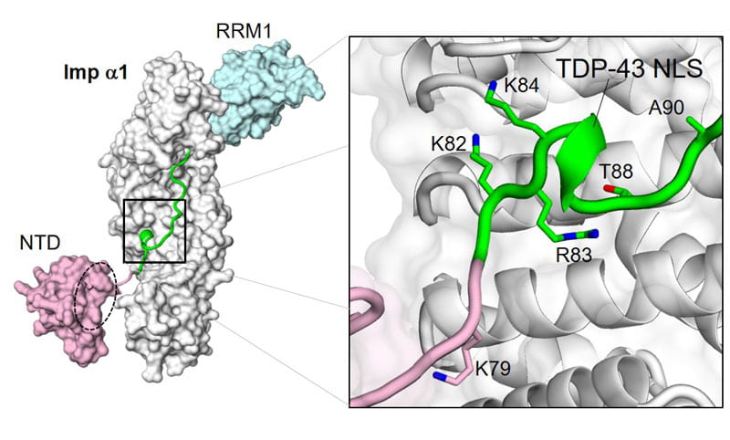
TDP-43 NLS bound to importin α1
PDB entry (X-ray): 7N9H
Doll et al., Cell Rep., 2022
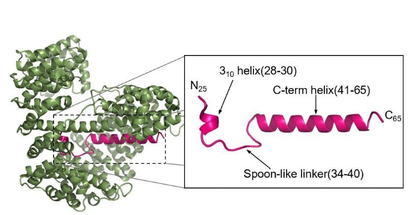
Pseudomonas-phage E217 TerS
PDB entries (Cryo-EM): 8DKR and 7UXE
Map: EMD-26858
Lokareddy et al., J Mol Biol., 2022
Pseudomonas-phage PaP3 Portal Protein
PDB entries (Cryo-EM): 7SXK, 7SYA, 7SZ4 and 7SZ6
Maps: EMD- 25500, 25521, 25560, 25562, and 25572
Chun-Feng David Hou et al., J Mol Biol., 2022
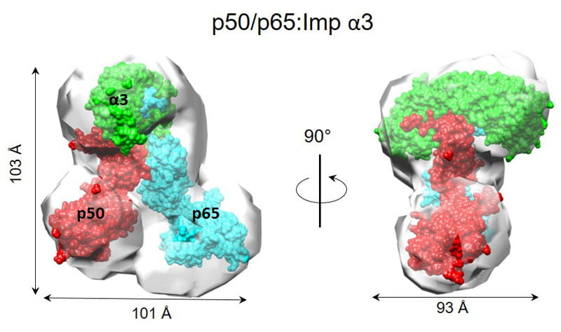
NF-kB p65/p50-Imp a3 Complex
PDB entries (X-ray/SAXS): 7LFC, 7LF4, 7LEU, 7LEQ, and 7LET
Florio et al., Nature Commun, 2022
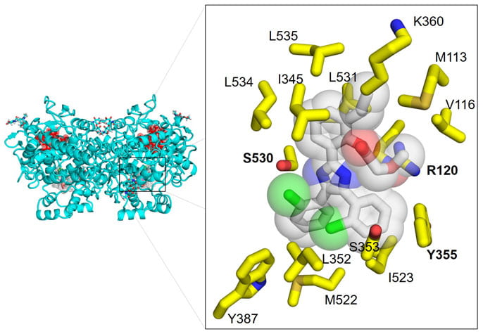
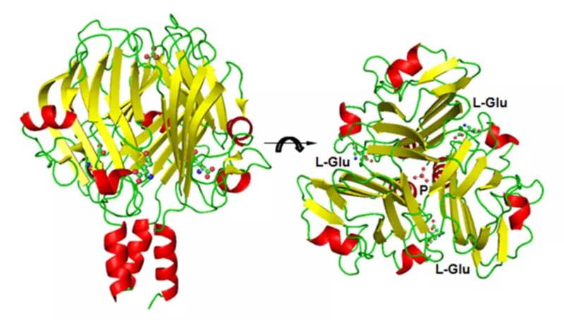
Synthetic COX-1 Inhibitor
PDB entry (X-ray): 7JXT
Friedrich et al., Adv Sci., 2021
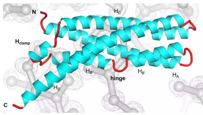
Cryo-EM structure of the T7 DNA-Ejectosome
PDB entry (Cryo-EM): 7K5C. Map: EMD-22680
Swanson et al., Molecular Cell, 2021
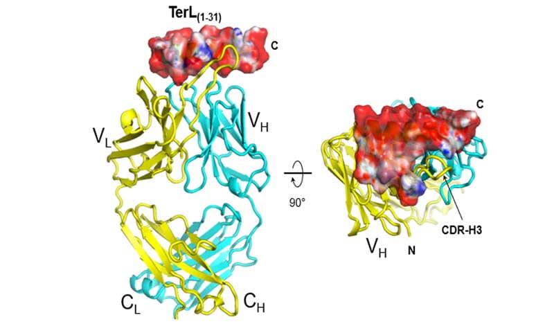
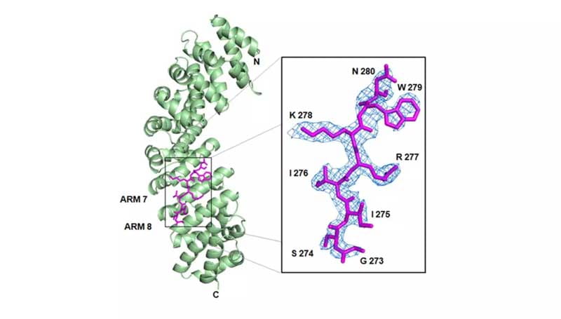
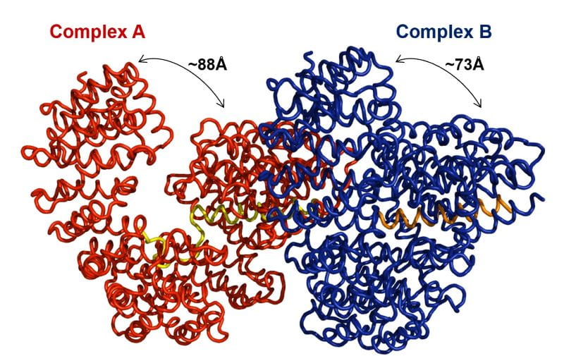
Recombinant Fab4 bound to TerL (1-31)
PDB entries (X-ray): 6VI2, 6VI1, 6XMI
Lokareddy et al., Acta D, 2020
Pseudomonas-phage PaP3 TerS
PDB entry (X-ray/SAXS): 6W7T
Niazi et al., Nucleic Acid Research, 2020
Chlamydia SNARE-like Protein IncA
PDB entries (X-ray): 6E7E, 6E6A
Cingolani et al., Nature Commun., 2019
Mycobacterium Tuberculosis Heme Transporter DppA
PDB entries (X-ray): 6E3D, 6E4D
Mitra et al., Nature Commun., 2019
Saccharomyces cerevisiae Inositol Phosphatase Siw14
PDB entry (X-ray/SAXS): 6E3B
Florio et al., Biochemistry, 2019
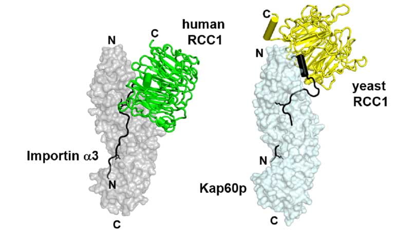
RCC1 bound to importin a3 and Kap60
PDB entries (X-ray/SAXS): 5TBK, 5T94
Sankhala et al., Nature Commun., 2017

Inhibition of COX-1 by Mofezolac and P6
PDB entries (X-ray): 5WBE, 5U6X
Cingolani et al., Eur. J. Med. Chem., 2017
Immature ProCapsid Portal Protein
PDB entries (X-ray): 5JJ3, 5JJ1
Lokareddy et al., Nature Commun., 2017
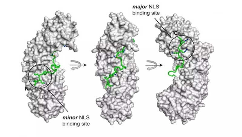
Pom121 NLS bound to importin α
PDB entry (X-ray): 4YI0
Kralt et al., Mol. Cell. Bio., 2015
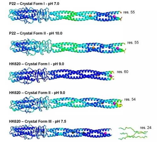
HK620 and P22 Tail Needles at different pHs
PDB entries (X-ray): 4ZKP, 4ZKU, 4ZXQ, 5BU5, 5BVZ, 5BU8
Bhardwaj et al., J. Biol. Chemistry, 2016

Heh1 and Heh2 membrane protein NLSs
PDB entries (X-ray): 4PVZ, 4XZR
Lokareddy et al., Structure, 2015
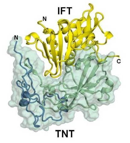
Mycobacterium Tuberculosis TNT-IFT complex
PDB entry (X-ray): 4QLP
Sun et al., Nature Struct. Mol. Biol., 2015
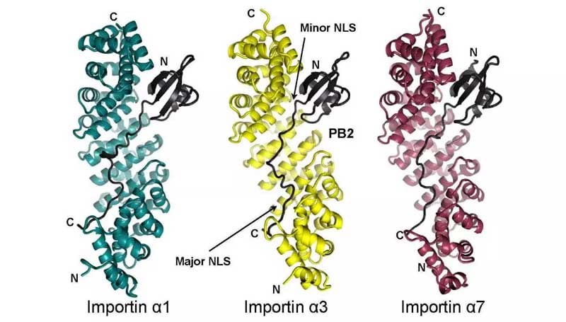
Importin α1, 3, 7 bound to Influenza PB2
PDB entries (X-ray): 4UAD, 4UAE, 4UAF
Pumroy et al., Structure, 2015
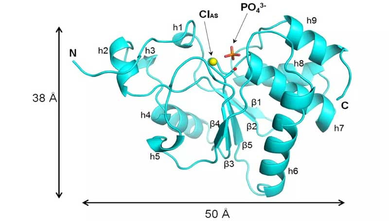
Phosphatase domain of Human Laforin
PDB entry (X-ray): 4R30
Sankhala et al., J. Biol. Chemistry, 2015
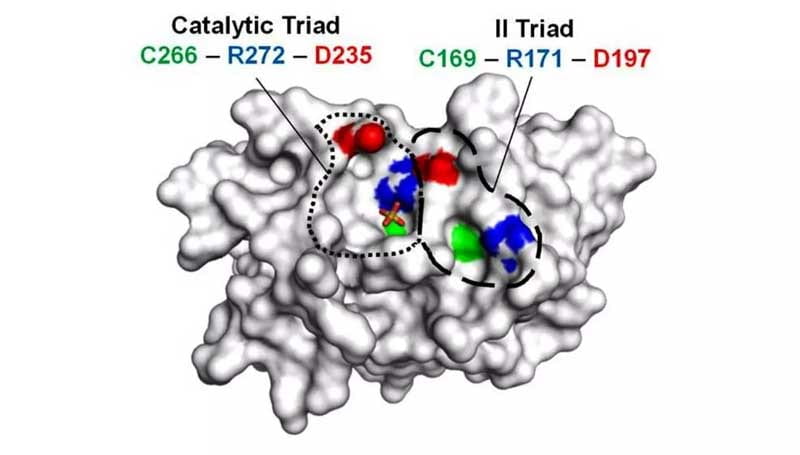
Human RNA Phosphatase PIR1
PDB entries (X-ray): 4MBB, 4NYH
Sankhala et al., Biochemistry, 2014

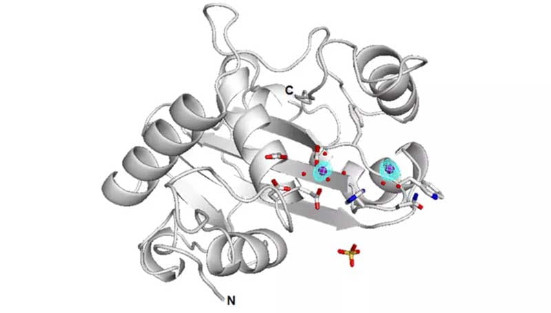
Human p53-phosphatase DUSP26
PDB entry (X-ray): 4HRF
Lokareddy et al., Biochemistry, 2013
P22 Headful Packaging Nuclease
PDB entry (X-ray): 4DKW
Roy et al., J. Biol. Chemistry, 2012
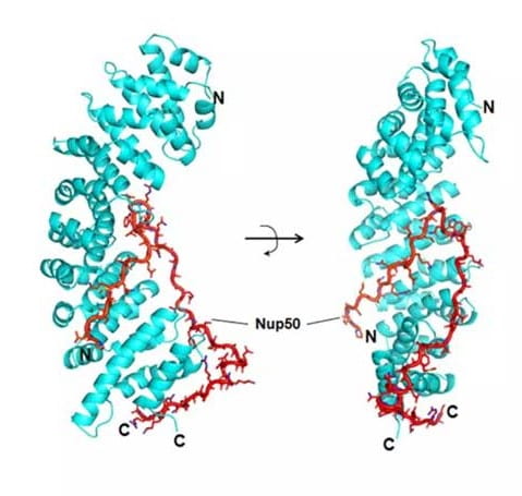
Human Importin α5 bound to Nup50
PDB entry (X-ray): 3TJ3
Ruth et al., J. Biol. Chemistry, 2012
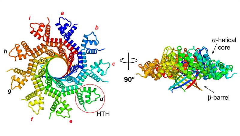
P22 nonameric Small terminase subunit
PDB entry (X-ray): 3P9A
Roy et al., Structure, 2012
Importin α1 bound to PLSCR4 minimal NLS
PDB entry (X-ray): 3Q5U
Lott et al., J. Biol. Chemistry, 2011
Structure of phage Sf6 Tail Needle Knob
PDB entries (X-ray): 3RWN, 4K6B
Bhardwaj et al., J. Biol. Chemistry, 2011
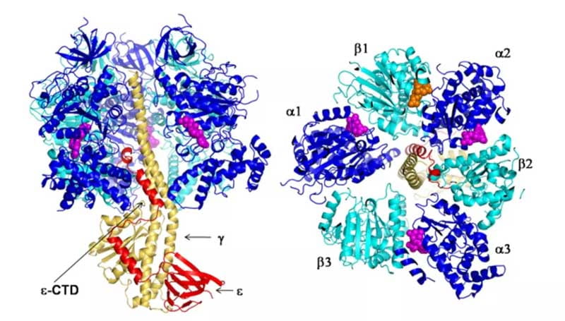
E.coli F1 ATPase inhibited by subunit ε
PDB entry (X-ray): 3OAA
Cingolani and Duncan, Nature Struct. Mol. Biol., 2011
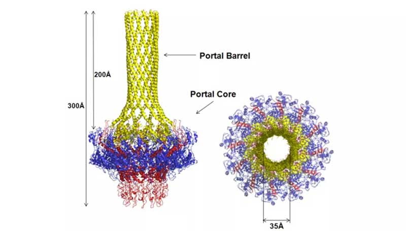
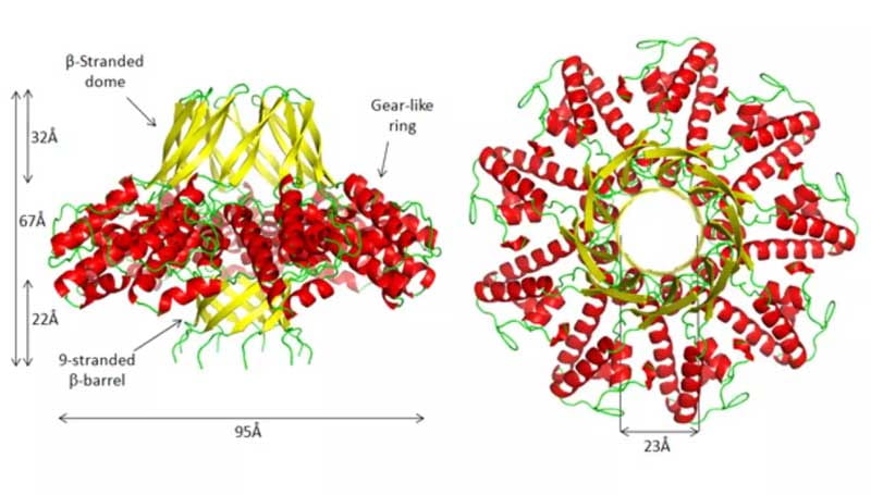
Full-length dodecameric P22 Portal Protein
PDB entry (X-ray): 3LJ5
Olia et al., Nature Struct. Mol. Biol., 2011
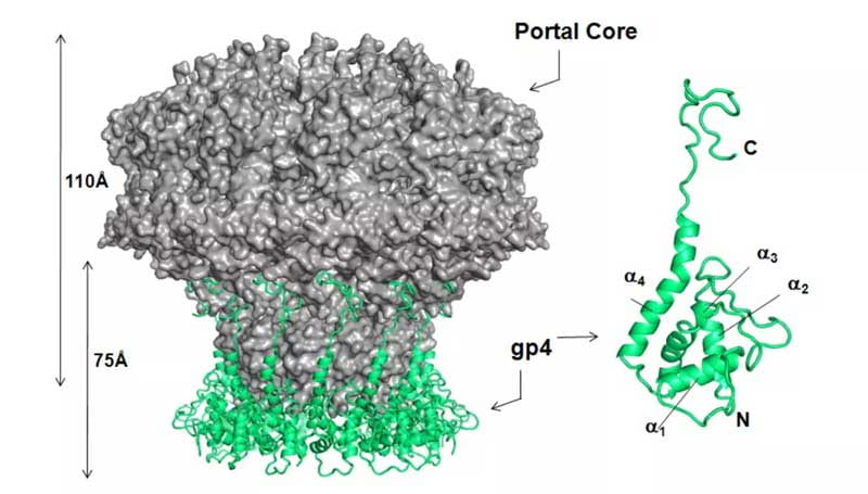
P22 Portal Protein Core bound to twelve GP4s
PDB entries (X-ray): 1VT0, 3LJ4
Olia et al., Nature Struct. Mol. Biol., 2011
Open and closed Importin β bound to sIBB
PDB entry (X-ray): 3LWW
Bhardwaj et al., Biochemistry, 2010
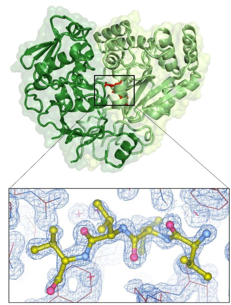
A circular permutant of Barnase
PDB entry (X-ray): 3DA7
Butler et al., Biochemistry, 2009
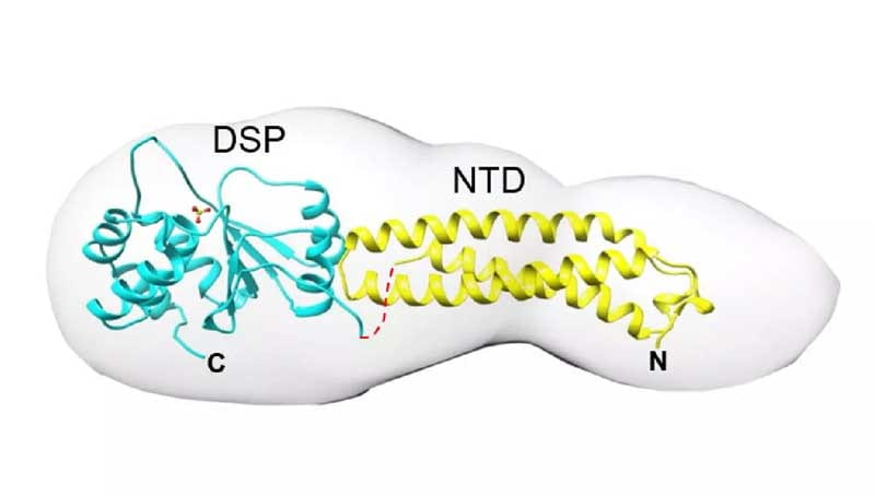
Vaccinia Virus Dual Specificity Phosphatase
PDB entries (X-ray/SAXS): 3CM3, 2RF6
Koksal et al., J. Biol. Chemistry, 2009
Phage P22 Tail Needle Gp26
PDB entries (X-ray): 2POH, 3C9I
Olia et al., Nature Struct. Mol. Biol., 2007
Importin β bound to snurportin IBB-domain
PDB entries (X-ray): 2P8Q, 2Q5D
Mitrousis et al., J. Biol. Chemistry, 2008

Mammalian Importin α bound to PLSCR1 NLS
PDB entry (X-ray): 1Y2A
Chen et al., J. Biol. Chemistry, 2005
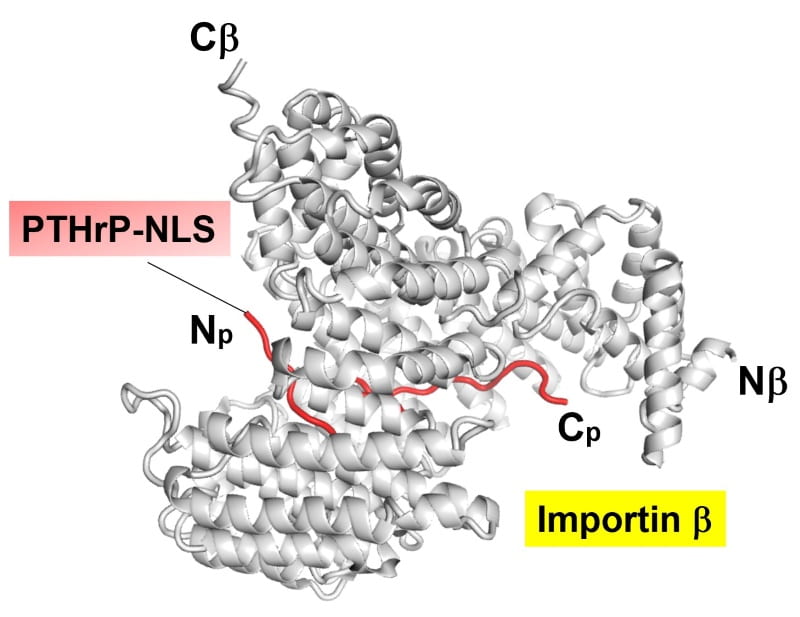
Importin β HEATs (1-11) bound to the PTHrP
PDB entry (X-ray): 1M5N
Cingolani et al., Molecular Cell, 2002
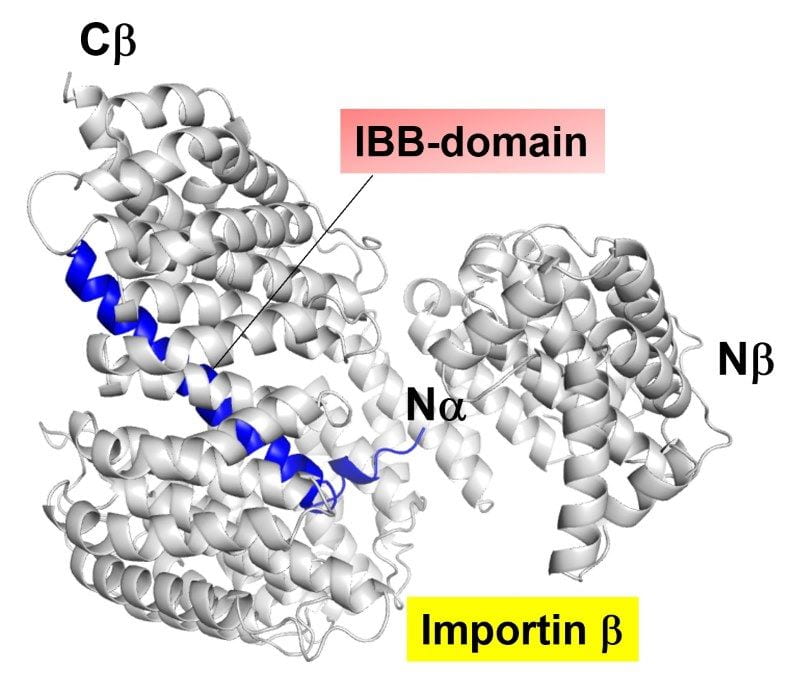
Human Importin β bound to the IBB-domain
PDB entries (X-ray): 1QGK, 1QGR
Cingolani et al., Nature, 1999
